
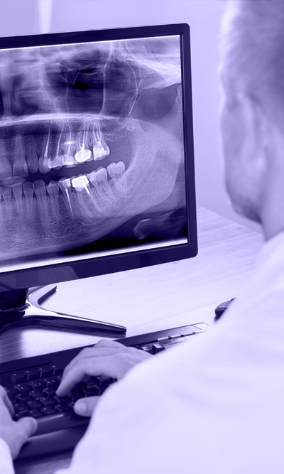
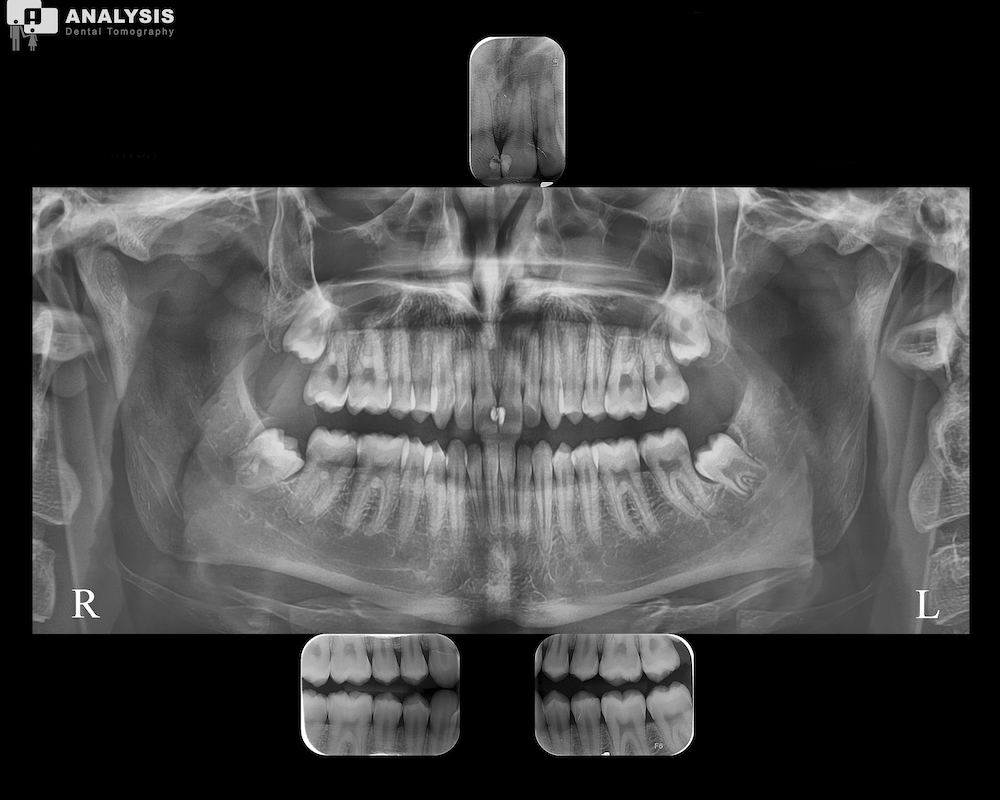
A panoramic radiograph is a two-dimensional image of the jaws, teeth, temporomandibular joints, and sinuses of the patient. It is often the reference point for assessing the condition of the teeth and jaws during the initial examination at the dentist. It is also useful for evaluating the progress of therapeutic interventions.
Panoramic X-ray is a very fast and safe examination. As it provides a complete image of the teeth and the bone that supports them in a few seconds, panoramic X-ray gives us information about the presence of pathology, such as tooth decay, periodontitis and the existence of damage to the jaw bone, as well as to the sinuses.
The panoramic radiograph, along with the lateral cephalometric radiograph, are the two initial examinations that the orthodontist will necessarily request when starting orthodontic treatment to assess the patient's needs and determine the stages of treatment. The panoramic radiograph is also necessary for the examination of the third molars (wisdom teeth), as well as their relationship with the neighboring teeth.
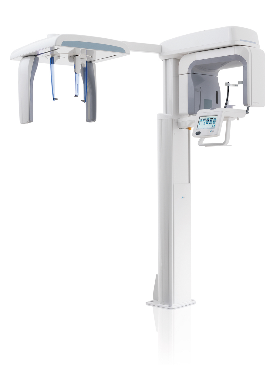
Digital technology allows us to make precise measurements and diagnoses using a computer. The transmission of the X-ray to the doctor who orders it is very fast and the results can be used immediately. Keeping records is also much easier with the use of digital technology, as digital X-rays can be stored on digital media and do not take up space. This way, the doctor has very easy access to the patient's history and can compare the radiological findings easily and quickly.

 English
  Greek
Greek