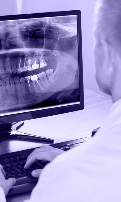
Retro-alveolar radiography of the teeth is a necessary examination in order to have a correct diagnosis, adequate documentation, and accurate planning of the treatment of the teeth and gums.
A proper retroalveolar radiograph must depict the tooth in its entirety, as well as the bone surrounding it, and even to an extent of 2 to 3 millimeters beyond the end of the tooth, the apical root.
To achieve a correct reproduction of the tooth image on the radiographic plate, there are two techniques: the bisection technique and the parallel imaging technique. There are advantages and disadvantages to both techniques, with the parallel imaging technique being preferred where possible.
Retro-alveolar radiography provides us with very accurate information, with the only limitation being that it is a two-dimensional image of a three-dimensional area, which means that it does not provide us with depth information. However, with some variations in the aiming technique, we can also have information about the third dimension.


 English
  Greek
Greek