
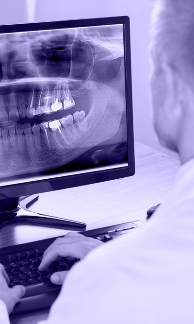
Perhaps the most important diagnostic tool that has been introduced in recent years in the field of dentistry is the introduction of cone beam volumetric imaging (CBVI) technology, which has however become commonly referred to as cone beam computed tomography (CBCT).
The common point between classic medical computed tomography (computed tomography CT or computed axial tomography CAT) and CBCT is the visualization of the final two-dimensional radiographic image.
However, there are also several differences, the most important of which is the amount of radiation the patient receives. For an examination of the jaw area using CBCT, the dose the patient receives can be up to 20 times lower than the dose required for the same examination using classic CT.
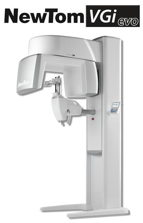
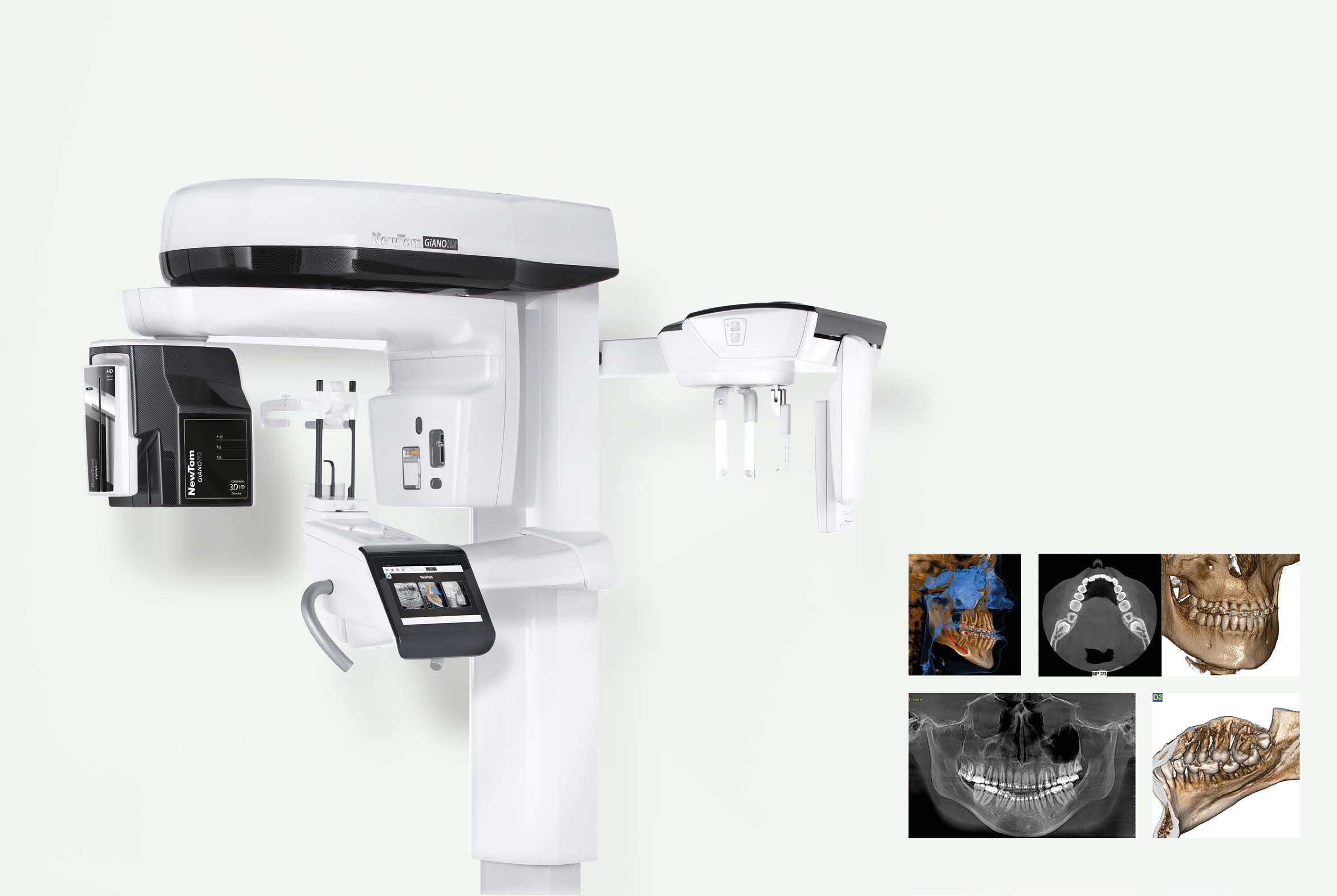
Also, the examination using classical CT is usually performed with the patient lying on the X-ray table. In contrast, in the CBCT examination, except in the case where we want to examine the airway, in which case it is advisable to
During a classic CT examination, the information is collected in pixel form and the depth element is added by the examiner, depending on the capabilities of each machine. This results in a volume element (voxel) with standardized but not predetermined dimensions, and the personal factor (the examiner's experience) also enters into play.
The exceptional accuracy and analysis of CBCT is so impressive that it has the effect of drastically influencing the treatment plan with the amount of information it provides. CBCT typically leads to more complete and effective treatment plans, helping the clinician make the right decisions. And as a communication tool with the patient, it offers the opportunity to explain and analyze the stages of treatment, helping the patient make the right decision.
Digital technology allows us to make precise measurements and diagnoses using a computer. The transmission of the X-ray to the doctor who orders it is very fast and the results can be used immediately. Keeping records is also much easier with the use of digital technology, as digital X-rays can be stored on digital media and do not take up space. This way, the doctor has very easy access to the patient's history and can compare the radiological findings easily and quickly.

 English
  Greek
Greek